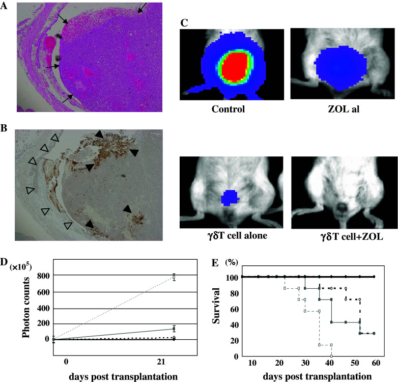
Fig. 4.
In vivo effects of intravesical administrated γδ T cells. After 3-h incubation with ZOL and γδ T cells, the bladders was occupied by a large tumor (↑) were dissected and examined histologically with hematoxylin-eosine staining (a) and immunohistochemically with anti-human CD3 antibody (b). Although the region of healthy bladder epithelium (triangle) was not infiltrated by γδ T cells, the tumor was massively infiltrated by γδ T cells indicated by human CD3 positivity (filled triangle). Original magnification ×16. The data shown are representative of 3 independent experiments. c In vivo effects of intravesical γδ T cells in the orthotopic bladder cancer murine model. Typical images of the respective mice not treated or treated with a low dose of ZOL alone, γδ T cells alone, or γδ T cells and a low dose of ZOL, were observed by IVIS. d The growth curves of orthotopically transplanted UM-UC-3LUC were measured by IVIS. The anti-cancerous effect of intravesical ex vivo-expanded γδ T cells was demonstrated in vivo: no treatment, open square; treatment with a low dose of ZOL alone, closed square; treatment with γδ T cells alone, open circle; treatment with γδ T cells and a low dose of ZOL, closed circle. e The survival curves of mice not treated or treated with γδ T cells alone, a low dose of ZOL alone, or γδ T cells and a low dose of ZOL. Survival of the orthotopic mice was improved by the intravesical administration of ex vivo-expanded γδ T cells: no treatment, open square; treatment with a low dose of ZOL alone, closed square; treatment with γδ T cells alone, open circle; treatment with γδ T cells and a low dose of ZOL, closed circle

