Fig. 1.
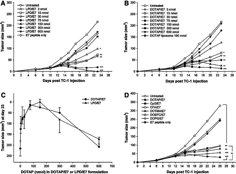
Kinetics of TC-1 tumor growth in mice treated with (a) LPD/E7 or (b) DOTAP/E7 formulation. a C57BL/6 female mice of 7 weeks old (n = 8–12) were injected sc with TC-1 tumor on day 0. On day 6, the mice received 10 μg E7 peptide formulated in LPD at various DOTAP lipid concentrations. The untreated mice were used as a negative control. b On day 6 post TC-1 inoculation, the mice received treatment of 10 μg E7 peptide formulated in DOTAP liposomes at various lipid concentrations. Tumor sizes were measured with calipers and determined by multiplying the two largest dimensions of the tumor. Tumor size of each group on day 23 was compared to the untreated control group and was analyzed statistically (*P < 0.05, **P < 0.01, ***P < 0.001). c The anti TC-1 tumor activity of the formulations DOTAP/E7 and LPD/E7 was contrasted on day 23 by comparing the tumor size at each corresponding DOTAP lipid concentration. No statistically significant differences in tumor size were found between the two groups. d Anti-cancer activity of DOTAP/E7 was compared to that of other cationic lipids (DOEPC and DOTMA), an anionic lipid (DOPG), CFA and CpG ODN1826. Tumor-bearing mice (n = 6–12) received a single treatment on day 6 after tumor inoculation. TC-1 tumor sizes were measured and analyzed statistically

