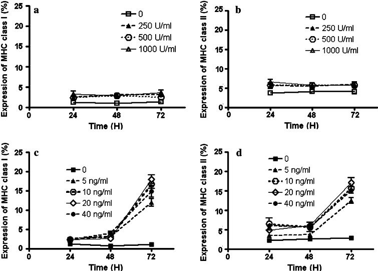
Fig. 5.
MHC class I and II antigen expressions on the P phase tumor cells stimulated by IFN-γ or IL-6. Isolated tumor cells from six tumor isolates were incubated with different concentrations of IFN-γ (250, 500, 1,000 U/ml) or IL-6 (5, 10, 20, 40 ng/ml) for 24, 48, or 72 h, and expression of MHC class I (a and c) and class II (b and d) antigens was determined by flow cytometry. None of the IFN-γ treatments increased MHC expression significantly. Different concentrations of IL-6 increased MHC class I and II antigen expressions, but only on tumor cells incubated with IL-6 for 72 h. Each experiment was done in triplicate

