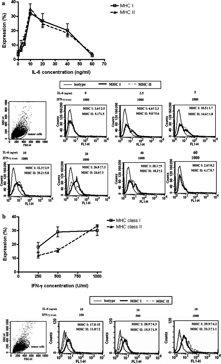
Fig. 7.
Expression of MHC class I and II antigens on tumor cells stimulated by different concentrations of IFN-γ/IL-6 when anti-TGF-β Ab is not present. Effect of 72 h exposures a IFN-γ (1,000 U/ml) and IL-6 (2.5, 5, 10, 20, 40, or 60 ng/ml), and b IFN-γ (250, 500, or 1,000 U/ml) and IL-6 (10 ng/ml) on the expression of MHC class I and II antigens in six tumor isolates were determined by flow cytometry. The upper portions of a, b displayed the expression curves of MHC I and II antigens stimulated by different concentrations of IL-6 or IFN-γ, respectively. Peak expression was achieved with 1,000 U/ml IFN-γ and 10 ng/ml IL-6 (P < 0.05). Nevertheless, IL-6 concentration > 10 ng/ml resulted in lower levels of MHC antigen expression. The lower portions of a and b displayed the histograms corresponding to a and b

