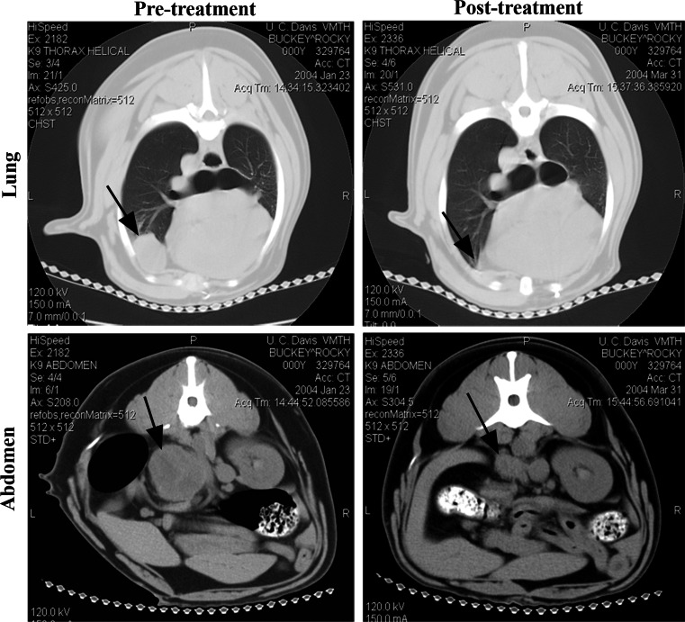
Fig. 7.
Tumor responses following LC treatment in a dog with malignant histiocytosis. A dog with metastatic MH was evaluated by CT imaging. Prior to treatment (left panels), there were large metastases in the lungs (upper panel, arrow) and the adrenal glands (bottom panel, arrow). The dog was then treated with two i.v. infusions of LC 2 weeks apart, as described in “Methods”. When CT imaging was repeated 3 months later (right panels), there was marked reduction in the size of both lung and adrenal metastases. At necropsy 5 months later, there was no evidence of MH in either the lung or adrenal sites

