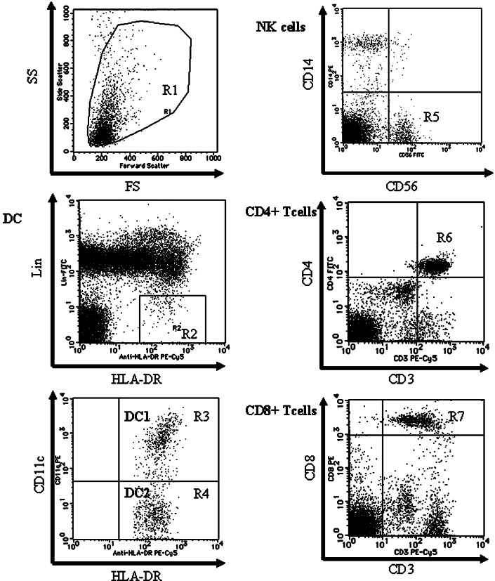
Fig. 1.
Flow cytometric analyses of PBMCs by FACS can. The figures are representative of a pancreatic cancer patient. Region R1 includes lymphocytes and monocytes but excludes debris. DCs are detected in region R2 as the population of Lin−/HLA-DR+ and divided into two fractions by the expression of CD11c (region R3; CD11c+ DC (DC1) and region R4; CD11c− DC (DC2)). The NK cell fraction is gated in the CD14−/CD56+ population (region R5). CD3+/CD4+ T lymphocytes are detected in region R6 and CD3+/CD8+ T lymphocytes are detected in region R7

