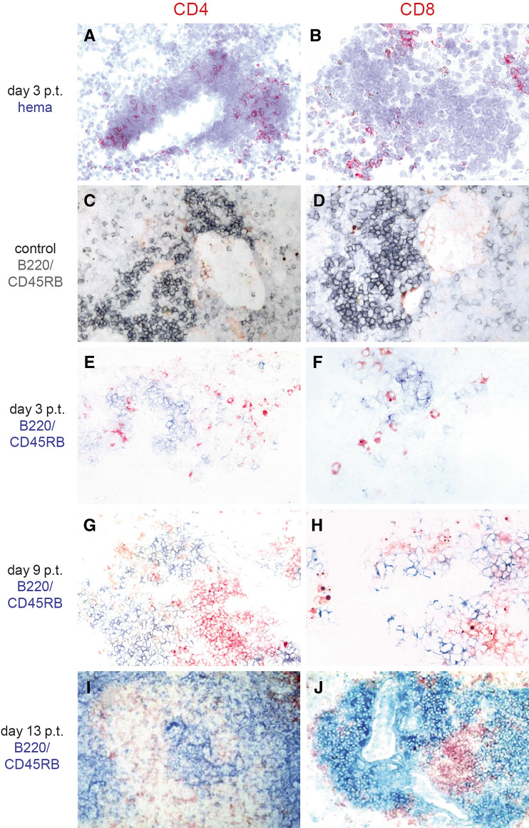
Fig. 3.
Immunhistological characterization of T-cell infiltrates. After i.v. (a, b, e–j) inoculation of 1.5 × 10⁶ B78-D14 cells, LTα−/− mice received 32 μg ch14.18-LTα fusion protein (a, b, e–j) for five consecutive days. Lungs were removed of a naïve control LTα−/− mice not receiving any therapy (c, d) or 3 days (a, b, e, f), 9 days (g, h), and 13 days (I, J) after cessation of therapy (p.t.), respectively. Sections thereof were stained for CD4 (a, c, e, g, i) or CD8 (b, d, f, h, j). In C to J sections were subjected to double staining with an anti-B220/CD45RB antibody (gray; c, d or blue; e–j) instead of counter-staining with hematoxylin (hema)

