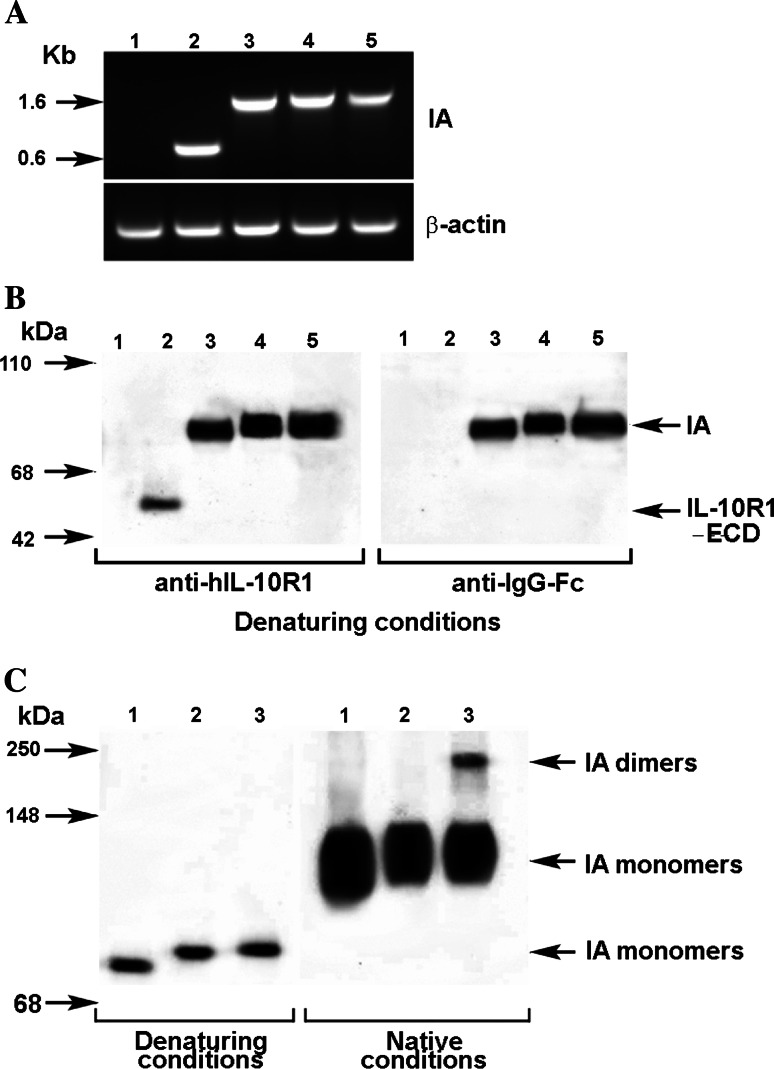
Fig. 2.
Detection of immunoadhesin. a mRNA derived from transfected pVAX1 vectors Total RNA was extracted from COS-1 cells that were transfected with immunoadhesins plasmid vectors. One-step RT-PCR was used to amplify the targeted mRNA. The PCR products were analyzed as described in Sect. Materials and methods. Immunoadhesins (IA): lane 1 #0 (wild type pVAX1), lane 2 #1 (IL10R1 extracellular domain only, 700 bps), lane 3 #2 (IL10R1/IgG1Fc with no hinge, 1,360 bps), lane 4 #3 (IL10R1/IgG1Fc with mutated hinge 1,400 bps), lane 5 #4 (IL10R1/IgG1Fc with wild type hinge 1,410 bps). β-actin (bottom) was served as an internal control to assure the integrity of mRNA. b Secretion of the immunoadhesin proteins. Serum-free culture media was collected 72 h after transfection of COS1 with different constructs. The culture media were then concentrated five times on Microcon YM50 filtration devise and separated on a 4–20% SDS PAGE followed by a transfer onto PVDF membranes in reducing (denaturing) conditions. Membranes were probed with either goat anti-human IL-10R1 antibody or with mouse anti-human IgG (Fc specific) antibody. Lane 1 #0 (backbone pVAX1), lane 2 #1 (IL10R1 ECD only), lane 3 #2 (IL10R1/IgG1Fc with no hinge), lane 4 #3 (IL10R1/IgG1Fc with mutated hinge), lane 5 #4 (IL10R1/IgG1Fc with wild type hinge). c Dimeralization of immunoadhesins. The concentrated culture supernatants obtained from transfected COS-1 cells were analyzed by the Western blotting method in reducing (denaturing) and non-reducing (native) conditions. Proteins were detected on blots by using anti-human IgG Fc specific antibodies. Dimerization of the immunoadhesin #4 (IA dimmers in lane 3) was confirmed in native conditions with the presence of higher molecular weight protein. Lane 1 #2 (IL10R1/IgG1Fc with no hinge), lane 2 #3 (IL10R1/IgG1Fc with mutated hinge), lane 3 #4 (IL10R1/IgG1Fc with wild type hinge)

