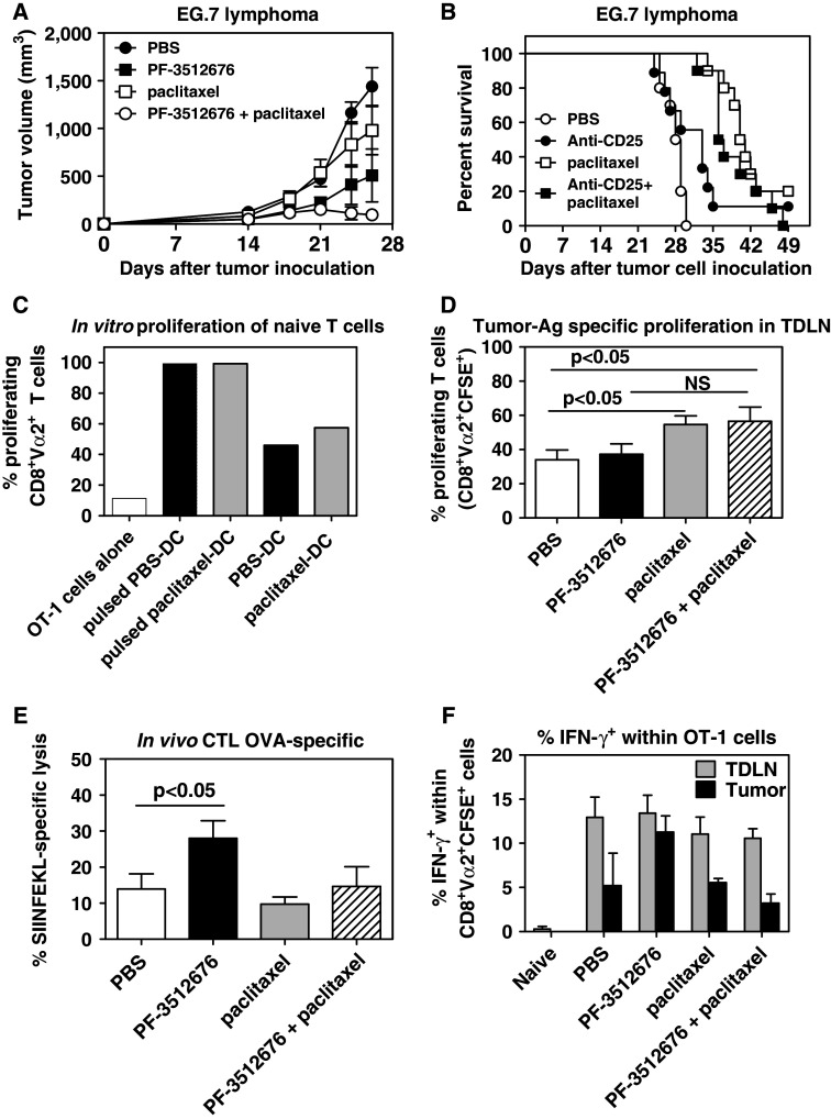
Fig. 4.
Paclitaxel increased the priming of tumor-antigen specific CD8 T cells but decreased the systemic frequency of tumor-antigen specific effector CD8 T cells. a Effect of weekly administration (D7, D14, D21) of PBS (closed circles), IP paclitaxel (open squares), peri-tumoral PF-3512676 (closed squares) or both (open circles) on tumor growth in the EG.7 tumor model. Results are representative of three independent experiments. b Effect of weekly administration (D7, D14, D21) of PBS (open circles), IP anti-CD25 (closed circles), IP paclitaxel (open squares) or both (closed squares) on tumor survival in the EG.7 tumor model. P < 0.001 for paclitaxel or paclitaxel plus anti-CD25 versus PBS; anti-CD25 versus PBS NS. c In vitro proliferation of OT-1 cells over 72 h, measured by CFSE staining dilution, in the presence of tumor-associated dendritic cells purified from mice treated with IP paclitaxel (gray bars) or PBS (black bars) 48 h before, and with (pulsed) or without additional OVA SIINFEKL peptide. White bars: OT-1 cells alone. Results are representative of two independent experiments. d Proliferation of adoptively transferred OT-1 cells, measured by CFSE staining dilution, in tumor draining lymph nodes from EG.7 tumor-bearing mice 3 days after transfer and 5 days after treatment with PBS (white bars), peritumoral PF-3512676 (black bars), IP paclitaxel (gray bars) or both (hatched bars). Results are cumulative of three independent experiments (n = 4–5 per experiment). e OVA SIINFEKL-specific in vivo CTL activity assessed in spleen as described in "Materials and methods" in EG.7 tumor-bearing mice 2 days after treatment with PBS (white bars), peritumoral PF-3512676 (black bars), IP paclitaxel (gray bars) or both (hatched bars). Results are representative of 3 independent experiments (n = 5 per experiment). f Intra-cellular IFN-γ expression within adoptively transferred OT-1 cells in EG.7 tumor draining lymph nodes (TDLN, gray bars) or tumors (black bars) 3 days after transfer and 5 days after treatment with PBS, peritumoral PF-3512676, IP paclitaxel or both. Expression in OT-1 cells transferred into naïve animals is also shown. Results are representative of two independent experiments (n = 5 per experiment). Statistical analyses: Mann–Whitney test

