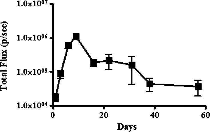
Fig. 1.
Development of localized osteomyelitis in the mouse. Mice (n = 5 per group) were challenged with luciferase-transfected Staphylococcus biofilm-coated suture material introduced into the medullary cavity of the tibia, and imaged using a Xenogen In Vivo Imaging System to track the intensity and localization of the infection. Time course analysis of infection intensity of osteomyelitis with means (±SEM) calculated

