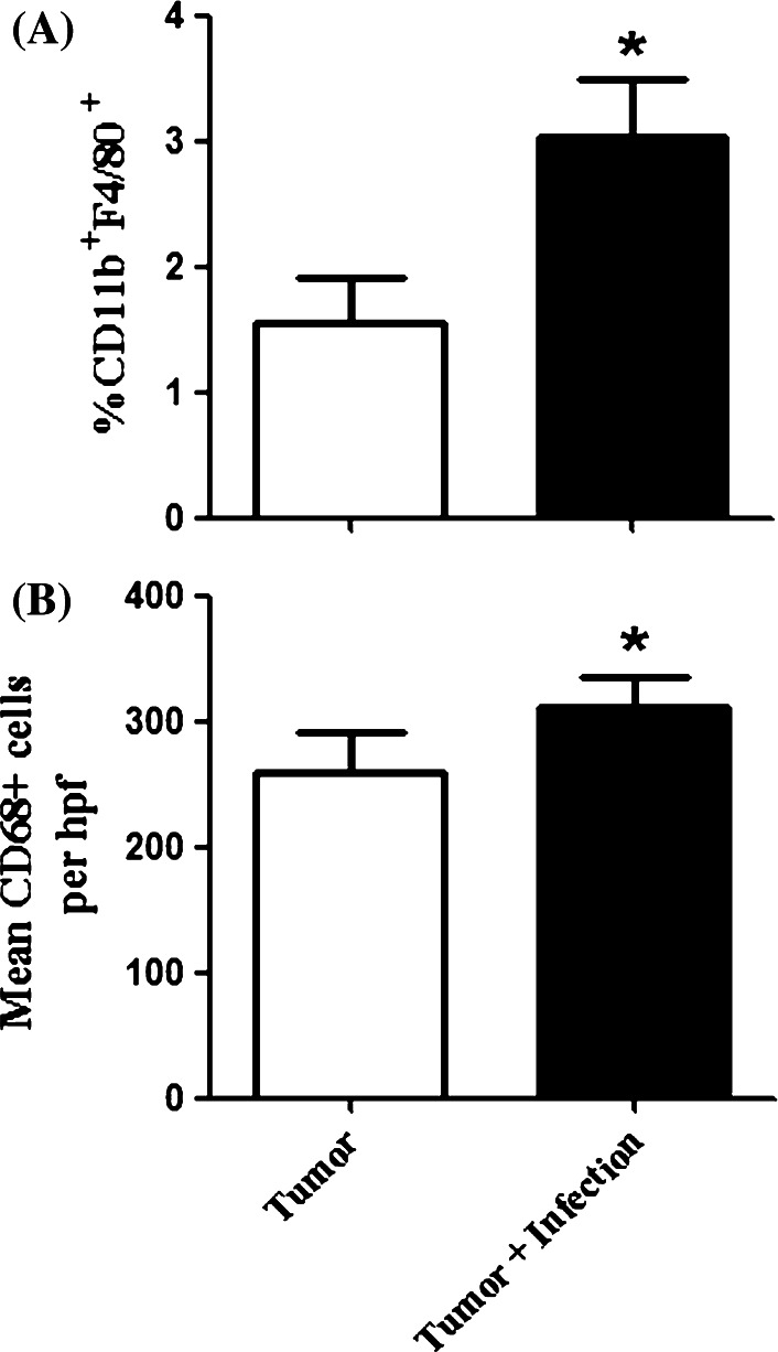
Fig. 6.
Macrophages are induced in infected, tumor-bearing mice. Tumor-bearing mice (n = 5 per group) were sacrificed when the first control mouse reached a maximal tumor diameter of 10 mm. Tumors were removed and homogenized for analysis by flow cytometry or prepared for IHC as described in the Materials and methods. a There was a significant increase (*p = 0.03) in the number of CD11b+F4/80+ macrophages in the tumors of infected mice compared to uninfected mice. b There was a significant increase in the number of CD68+ cells per 20× high power field (hpf) in the tumor tissue of infected mice compared to the uninfected mice (*p = 0.019). Results are representative of two independent experiments and two-tailed t tests were used to determine significance

