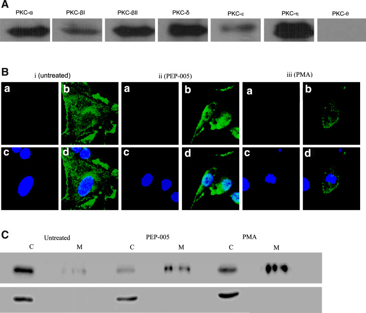
Fig. 4.
PEP005 or PMA mobilise PKC-δ to the perinuclear membrane. a EC were assessed for classical and novel PKC isoform content by western blotting and found to express high levels of six isoforms. b EC were assessed for PKC-δ activation by immuno-fluorescent staining and confocal microscopy in i untreated cells or cells treated with either ii PEP005 or iii PMA. a Shows cells stained with an irrelevant isotype matched antibody; b with anti-PKC-δ antibody; c with the nuclear stain DAPI; d a merged image of b and c. c EC were assessed for translocation of PKC-δ by western blot after extraction of cytoplasmic (C) and membrane (M) associated fractions after no treatment (untreated) or treatment with PEP005 or PMA. Upper panel shows PKC-δ immunoreactivity and the lower panel is the loading control β-actin. Both means of assessment showed that PKC-δ was translocated from a cytoplasmic to membrane location in response to PEP005 or PMA

