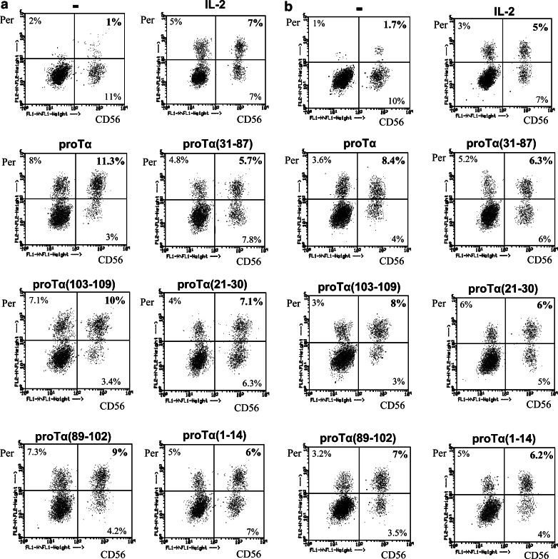
Fig. 3.
Dot plot diagrams of 3-day cultured healthy donor (a) and cancer patient (b) PBMC with IL-2 (20 IU/ml) and proTα or its tryptic fragments. PBMC were labeled with anti-CD56-FITC (CD56)- and anti-perforin-PE (Per)-specific antibodies. Percentages of double positive cells refer to the lymphocyte population gated. Data are from one representative experiment out of 3 and 2, conducted for healthy donors and cancer patients, respectively— PBMC incubated with medium; IL-2, PBMC incubated with 20 IU/ml IL-2 alone

