Fig. 1.
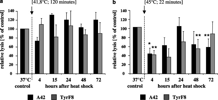
Susceptibility of heat-treated 624.38-MEL melanoma cells to CTL-mediated killing after heat exposure. Effector CTLs were TyrF8 and A42, which recognize the tyrosinase or MART-1/MelanA peptide in an HLA-A2-restricted manner [16]. Target cells were the melanoma cell 624.38-MEL exposed to two different thermal doses pre-determined by clonogenic survival assays to result in similar cell survival (isosurvival dosis) [16]. Shown in (a) are the relative lysis values of 624.38-MEL after exposure to low temperature and long duration (41.8°C/120 min) and in (b) after high temperature and short duration (45°C/22 min) expressed as percentage compared to control cells at 37°C. Cells were exposed to heat treatment by submerging the sealed flask into a temperature-controlled water bath. Control flasks were sealed and left at 37°C. After heat exposure flasks were returned to 37°C and humidified at 5% CO2 atmosphere. At indicated time-points viable cells were harvested, labeled with 51Cr for 1 h and co-incubated with titered effector CTL, TyrF8 and A42, at a constant cell number of 2,000 cells per well in 96-V bottom plates. Duplicate measurements of four-step titrations of effector were used in all experiments. Spontaneous and maximum releases were determined by incubating the target cells alone and by directly counting labeled cells, respectively. After 4 h of incubation at 37°C in a humidified 5% CO2 atmosphere, supernatants were harvested, transferred to Lumaplate solid scintillation microplates, dried over night and counted on a TopCount microplate scintillation counter (Packard, Meriden, CT, USA). For each E:T ratio, the percentage of lysis was calculated as follows: % specific lysis = (experimental cpm − spontaneous cpm/maximal cpm − spontaneous cpm)×100. The summarized results (±STD) of two independent experiments obtained at an effector to target ratio of 10:1 are shown. Relative lysis values were calculated as the percentage of specific lysis compared to control cells at 37°C which was set to 100%. Absolute values of specific lysis at 37°C were 19%±5 for A42 and 34%±12 for TyrF8. The statistical significance of experimental values was assessed by means of independent Student’s t test comparing melanoma cells at 37°C and after heat-shock treatment. P values of P<0.05 (*) are significant, P< 0.01 (**) are considered as highly significant

