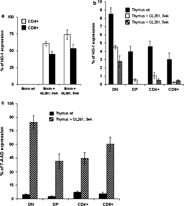
Fig. 5.
Apoptosis of T-cells of glioma bearing mice: a flow cytometry analysis of gated CD4+ and CD8+ cells infiltrating brain of glioma bearing mice (3 and 5 week) compared to littermate brain wt control, stained for CD4, CD8 and HO-1. The result was recapitulated in a histogram; b expression of HO-1 in gated thymocytes DN, DP, SP CD4+ and SP CD8+ analyzed with flow cytometry and represented in a histogram; c representative histogram of 7-AAD uptake detected by flow cytometry in different thymocytes subsets (DN, DP and SP) stained for CD4, CD8 and 7-AAD. Values represent the mean fluorescence intensity ± SD

