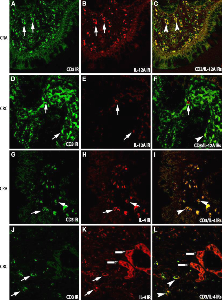
Fig. 3.
Double immunofluorescence staining for the examination of cytokine-producing lymphocytes in the tissues of adenoma–carcinoma sequence. In CRA, the increased numbers of lymphocytes labeled by CD3 immunorecativity (IR) (FITC, green; arrows a) were paralleled to the increased IL-12 expressing cells (TRITC, red) in the stroma (arrows b), but not in carcinoma sections (CRC, e), in which IL-12 IR cells were remarkably decreased although CD-3 IR cells were increased; Consequently, the cell number with co-localization of CD3/IL-12 IRs in CRC (f) were remarkably lower than that in CRA (arrow heads c). The IR of Th2 cytokine IL-4 (TRITC, red) could be detected in both CRA and CRC (h, k). However, the IR of IL-4 was not only detected in the stroma cells (arrows k), but also in malignant epithelium (fingers point k, l) as compared with that in CRA (h). (Double immunofluorescence staining, original magnification ×400)

