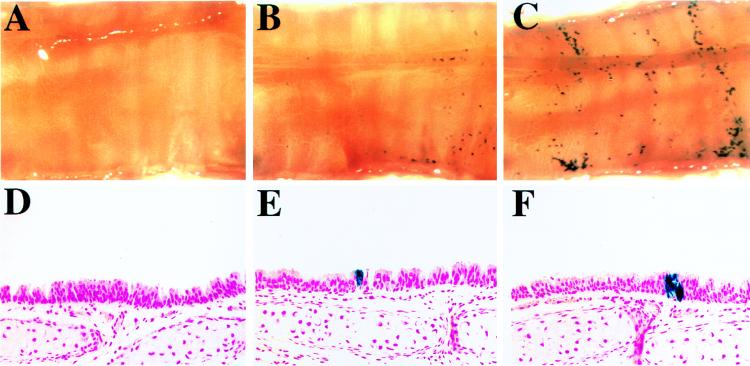
FIG. 10.
In vivo gene transfer to tracheas of mice 5 weeks of age. (A) En face view of an X-Gal-stained control (sham-infected) trachea opened longitudinally. (B and C) En face views of two X-Gal-stained mouse tracheas infected in vivo with concentrated HIT-LZ. (D to F) Representative histologic sections (counterstained with nuclear fast red; magnification, ×50) taken from the control trachea shown in panel A (D) and HIT-LZ-infected tracheas shown in panels B and C (E and F).

