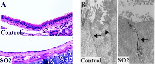
FIG. 6.
Morphologic assessment of SO2 injury. (A) Histologic sections of proximal air-exposed (control; upper panel) and SO2-exposed (lower panel) murine tracheas 24 h after inhalation of air or oxidant (stained with hematoxylin and eosin). (B) Lanthanum permeation (black staining) into the intercellular spaces (arrows) of tracheas from mice exposed to SO2 inhalation 24 h prior to sacrifice (right) compared to absence of lanthanum permeation into intercellular spaces of tracheas from mice sham-exposed to air (control) 24 h prior to sacrifice (left). Arrows depict the intercellular space. Magnification, ×3,000.

