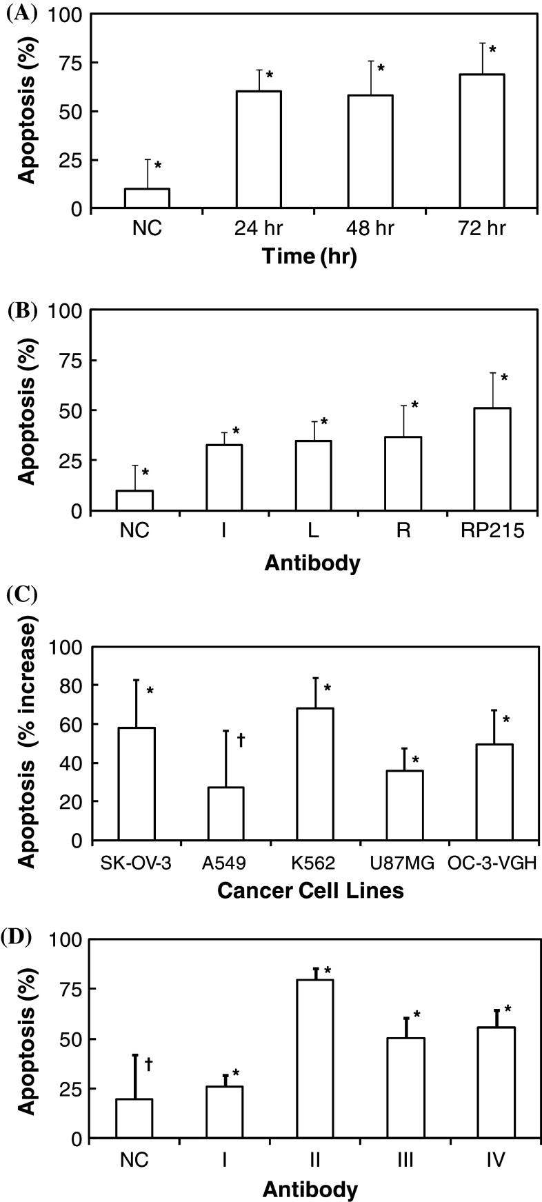
Fig. 3.
Percentage of cells with apoptosis of a OC-3-VGH ovarian cancer cells in culture upon incubations of RP215 (10 μg/ml) for 24, 48, and 72 h, respectively (all data revealed a statistical significance at p < 0.05); b OC-3-VGH ovarian cancer cells in culture upon incubations of RP215 (10 μg/ml) and anti-anti-id sera (Ab3) from three immunized mice designated as I, L, and R, respectively, at 1:1,000 dilution for 72 h (data are statistically significant at p < 0.05); c SK-OV-3, A549, K562, U87MG, and OC-3-VGH cancer cells in culture upon incubations with RP215 (10 μg/ml) for 24 h (*p < 0.05; † p > 0.05); data are presented after negative control is subtracted from the samples; and d OC-3-VGH ovarian cancer cells upon 48 h incubation with different antibodies including: I human IgG (10 μg/ml); II goat anti-human IgG (10 μg/ml); III RP215 (10 μg/ml); and IV RP215 (20 μg/ml). Data presented are percentage of cells with apoptosis. The negative controls were from cells upon incubation either with normal mouse IgG or serum of the same concentrations or dilutions (*p < 0.05; † p > 0.05). Standard deviations of each set of experiments in triplicates are presented by error bars (*p < 0.05; † p > 0.05) with means. ANOVA tests were performed for statistical significance defined at p < 0.05 for a–d

