
Fig. 5.
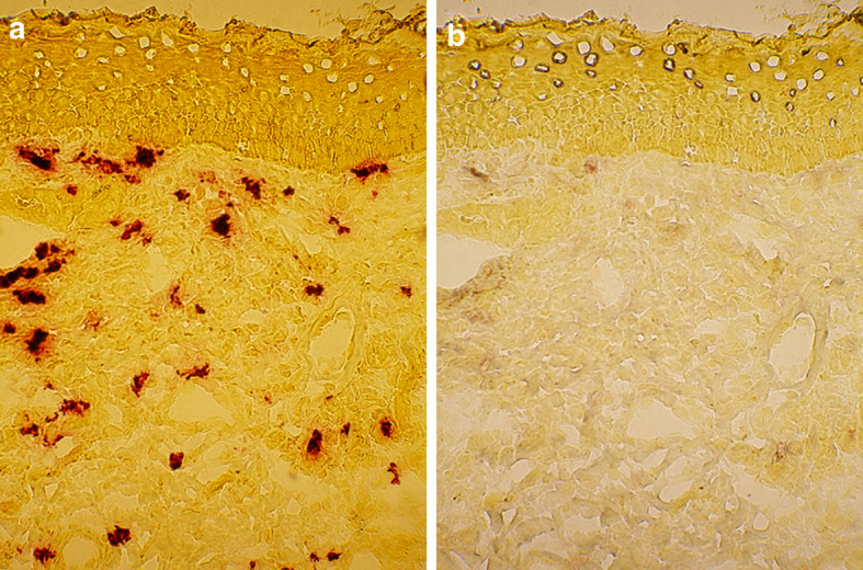
Mast cells do not display CD40L immunoreactivity in the BCC lesion. The lesional BCC section was first stained a enzymehistochemically for tryptase. After photographing, the red azo stain was dissolved away and the same section was stained b immunohistochemically for CD40L. Note that only rare occasional mast cells show weak CD40L immunoreactivity