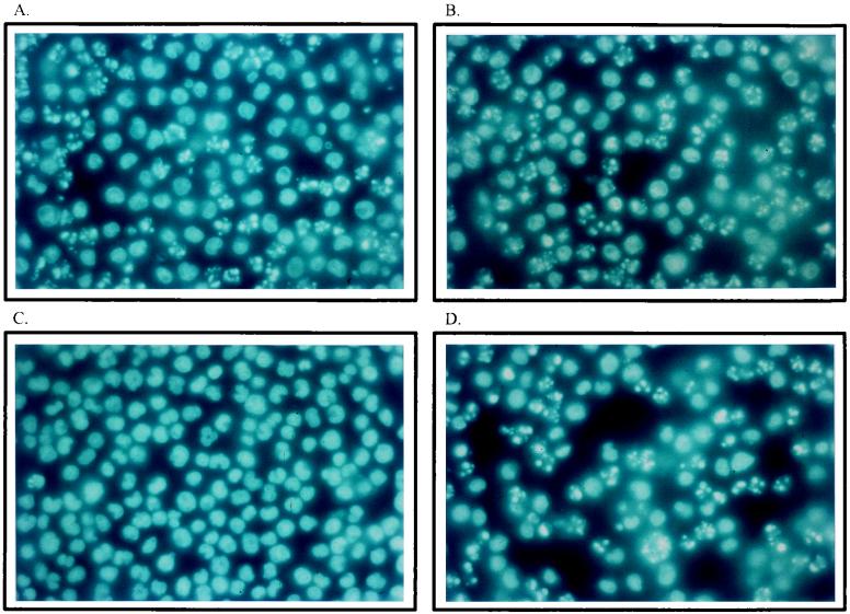
FIG. 1.
Induction of morphologic changes in U937 cells infected with H-1 virus. Cultures (106 cells) were infected with H-1 virus (5 PFU/cell) (A and B), mock infected (C), or treated with TNF-α (10 ng/ml) (D) and further incubated for 15 h (A) and 25 h (B, C, and D). Cells were collected by low-speed centrifugation and suspended in PBS before being fixed in 4% formalin on poly-l-lysine-coated slides, stained with Hoechst solution, and examined by fluorescence microscopy.

