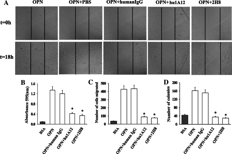
Fig. 4.
The inhibition effects of hu1A12 on MDA-MB-435S cell migration, adhesion, invasion and colony formation in soft agar. a Cell migration was inhibited by hu1A12. After wounding, MDA-MB-435S monolayer cells were cultured in medium containing OPN in the presence of 25 μg/ml of hu1A12 or control human IgG. Photographs were taken immediately or 18 h after wounding under a Carl Zeiss Axiovert 135 microscope (×40). The data represent one of three experiments with similar results. b hu1A12 inhibits cell adhesion to OPN. MDA-MB-435S cells with 25 μg/ml of hu1A12 or control human IgG were added to 96-well plates pre-coated with OPN. After 2 h incubation, unattached cells were washed off and the adherent cells were fixed in 1% methanol, stained with 0.5% crystal violet and lysed with 2% Triton X-100. The absorbance was measured at 595 nm. Columns mean (n = 3), bars SD; *P < 0.05 versus control human IgG. c Cell invasion was inhibited by hu1A12. Cell invasion assay was performed using a 24-well Transwell system as described in “Materials and methods”. Finally, the cells invading through Matrigel were stained with crystal violet and counted. Columns mean (n = 3), bars SD; *P < 0.05 versus control human IgG. d Hu1A12 inhibits the ability of MDA-MB-435S cell to form colonies in soft agar. MDA-MB-435S cells suspended in 0.3% agar solution were plated onto a layer of 0.5% agar solution in 24-well culture plates, followed by the addition of medium containing 25 μg/ml of hu1A12 or control human IgG every other day. Ten days later, the colonies containing at least 50 cells were counted. Columns mean (n = 3), bars SD; *P < 0.05 versus control human IgG. 2H8, a mouse anti-OPN monoclonal antibody specific for RGD domain of OPN, was used as positive control for wound migration, invasion, adhesion, colony formation assays

