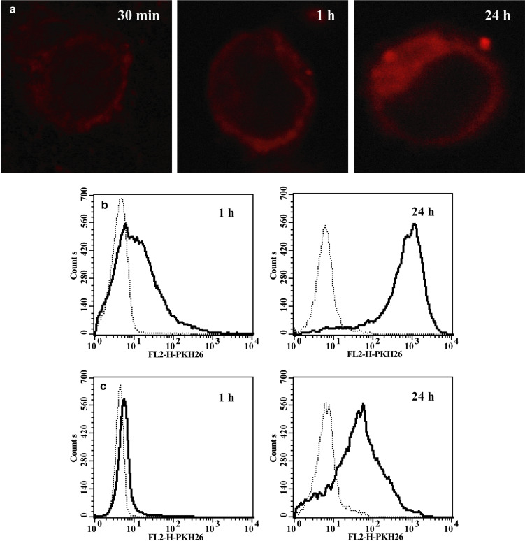
Fig. 4.
Transfer of PKH26 labelled TMVHPC to monocytes. a Confocal microscopy of monocytes exposed to TMVHPC for 30 min, 1 h and 24 h. b Flow cytometry of monocytes exposed to TMVHPC for 1 h and 24 h in the absence and presence of crystal violet. c Monocytes were incubated either in the medium (dotted line) or with TMV (30 μg/ml) (bold line)

