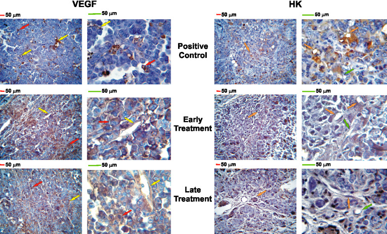
Fig. 5.
VEGF and HK immunohistochemistry: magnification: left ×400, right ×1000; red and green bars= 50 micrometers; positive staining is indicated by brown pigment within cytoplasm. VEGF (left panel): red arrows point to malignant tumor cells; yellow arrows point to stromal tissue (connective tissue, endothelial cells); The positive control group showed 5.2±0.9% of tumor and stromal cells positive to VEGF while both treatment groups showed more than 90% immunopositivity. HK (right panel): orange arrows point to HK-positive endothelial cells; green arrows point to HK-negative endothelial cells. The positive control group showed more than 75% of endothelial cell immunopositivity while both treatment groups showed significantly less HK-positive endothelial cells (early treatment: 27.2±1.8%; late treatment: 42.3±7.4%). There was no difference in immunopositivity for uPAR, FGF-2, BIR, and B2R among all experimental groups

