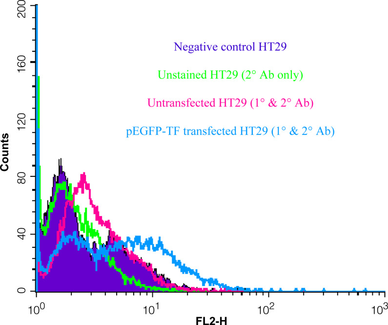
Fig. 1.
Flow cytometric assessment of TF antigen on the surface of HT29 cells. HT29 cells were transfected with pEGFP-TF and allowed to express the hybrid protein over 72 h. The cells were harvested and labelled with a mouse anti-human TF antibody. The cells were then washed and stained with a phycoerythrin-conjugated secondary antibody. The analysis of transfected and un-transfected cells was carried out by flow cytometry. A marker was set containing 5% of the control sample; 20% of untransfected cells and 49% for pEGFP-TF-transfected cells were in this region. Mean cell fluorescence intensity for the two samples were 3.7 and 7.5, respectively. The data is representative of three independent experiments

