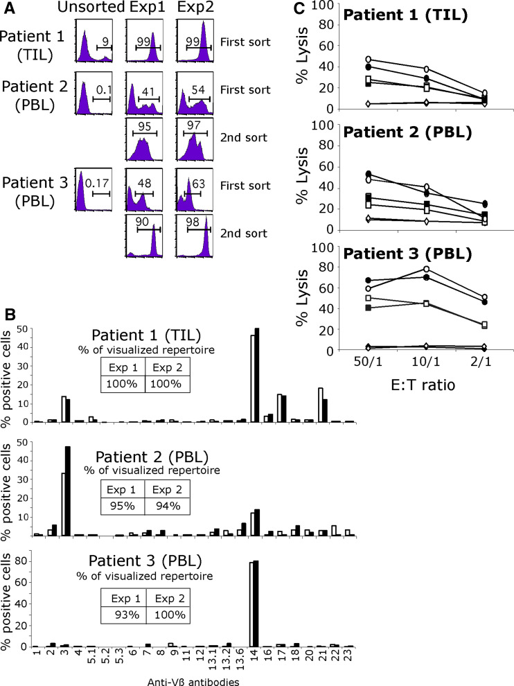
Fig. 1.
a Sorting of Melan-A specific lymphocytes from TIL and PBMC. HLA-A*0201/Melan-A-A27L monomers coated on magnetic beads were used to isolate in duplicate Melan-A-specific populations from melanoma patients’ PBMC or TIL. PE-conjugated tetramers were used to assess the purity of the sorted populations after a 14-day amplification on feeder cells. Values indicate the percentage of cells labeled with tetramers after one or two rounds of sorting/amplification. b Repertoire diversity of sorted populations was assessed by labeling with 24 anti-Vβ antibodies. Inserts indicate the percentage of the specific population characterized with this panel in each experiment: white bars experiment 1, black bars experiment 2. c Lysis of melanoma cell lines by Melan-A sorted populations: white and black symbols correspond to duplicate experiments. Circles and squares represent two HLA-A2 melanoma cell lines expressing the Melan-A antigen and lozenge a HLA-A2 negative melanoma cell line

