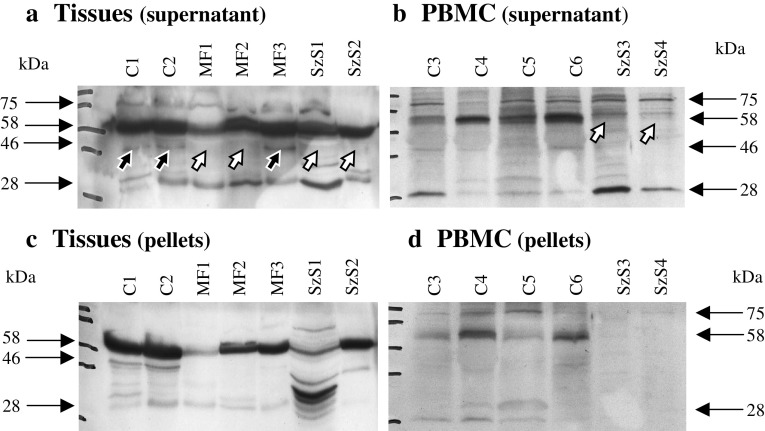
Fig. 4.
MT5-MMP and its variants in CTCL. Cells and tissues were mechanically disrupted and supernatant and pellet were analyzed separately. a Specimen of control skin (C1 and C2), three MF and two SzS specimen displayed the active 58 kDa form of MT5-MMP. The 46 kDa form is missing in several tumor specimens (open arrows) but present in controls and one MF specimen (closed arrows). b PBMC samples of four controls (C3 to C6) and two SzS patients differ in the presence or absence of the active 58 kDa form. Panels c and d represent the corresponding pellet fractions

