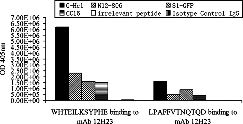
Fig. 2.
ELISA analysis of the phage competitive binding assay. M13 phage displaying peptides (WHTEILKSYPHE and LPAFFVTNQTQD) bound to mAb 12H23 were competed with different competitors. The competitors included Glycine–HCl (solid bars), N12-806 (right oblique bars), S1-GFP (left oblique bars), the CC16 peptide (horizontally hatched bars), irrelevant peptide (vertically hatched bars) and the isotype-matched control antibody (blank bars), respectively

