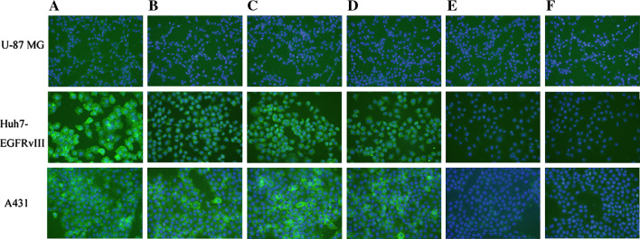
Fig. 5.
Immunofluorescence staining of EGFRvIII, EGFR overexpressed and normal EGFR expressed in Huh7-EGFRvIII, A431 and U-87 MG cells. Cells were incubated with anti-EGFR antibodies followed by FITC-labeled secondary antibodies where the nuclei were stained with DAPI. Cells were visualized with a fluorescence microscope. a Ch806 (positive control), b mAb 12H23 (positive control), c sera from mice immunized with WHTEILKSYPHE-KLH, d sera from mice immunized with LPAFFVTNQTQD-KLH, e sera from mice immunized with the control peptide-KLH, f sera from mice immunized with KLH alone

