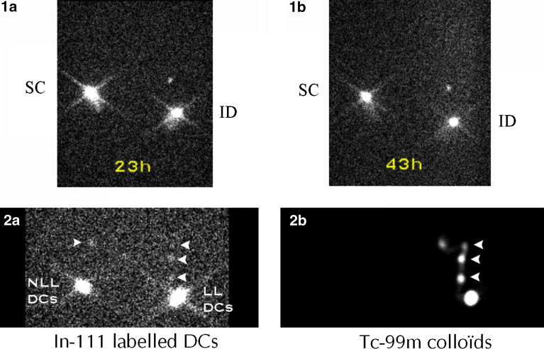
Fig. 1.
Migratory capacities of IL3/IFNβ DC. 1a, b In-111 labeled autologous IL3/IFNβ DC were injected in the proximal inguinal region of each leg of a patient, either subcutaneously (SC) on one side, or intradermally (ID) on the opposite side. Using a Sopha DSX gamma camera, 1a and 1b images, acquired, 23 h and 43 h post-injection, respectively, showed migration only with ID injection. 2a An image obtained 15.5 h after ID administration of labeled DCs either loaded (LL) with NA17.A2 antigen on one side, or non loaded (NLL) on the opposite side, demonstrated tracer migration towards nodular inguinal sites (arrow heads). 2b The patient was secondarily injected with Tc-99m nanocolloïds on the site of LL DCs injection. The acquisition obtained 30 min later demonstrated that the migration detected on In-111 2a image corresponded to three draining lymph nodes in the inguinal region

