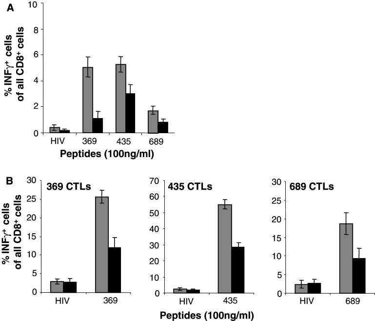
Fig. 6.
Impaired presentation by MC-2/HER-2 cells of HLA-A2 restricted epitopes. MC-2 (grey square) or MC-2/HER-2 (filled square) cells were pre-pulsed for 1 h at 37°C with control HIV or relevant HER-2 peptides and subsequently used to stimulate for 4 h ex vivo isolated (a) or 5 days in vitro peptide-re-stimulated splenocytes (b) from Ub/HER-2 mini-gene immunized HHD mice. The activated CD8+ T cells were quantified by intracellular cytokine staining for IFN-γ and analyzed by flow cytometry. Mean ± SD of three independent experiments is shown

