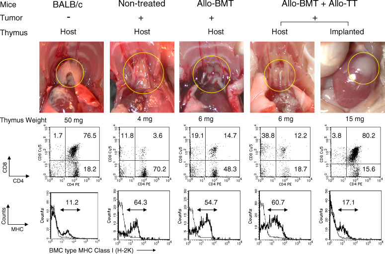
Fig. 3.
Findings related to host and transplanted thymus, CD4/CD8 subsets, and MHC expression in thymocytes from mice with advanced tumors treated with BMT + TT. The macroscopic findings (upper panel thymus in yellow circle), FACS profile (middle panel), and MHC class I (H-2K) expression (lower panel) in thymocytes from the host and transplanted thymus in BALB/c mice, non-treated controls with advanced tumors, and those treated with Allo-BMT and Allo-BMT + TT are shown. Autopsy and analysis were performed at the same time as those in Fig. 2. H-2K expression thick line, BMC type (H-2Kd in BALB/c and non-treated controls, H-2Kb in Allo-BMT and Allo-BMT + Allo-TT), thin line, negative control or host type (H-2Kb in BALB/c and non-treated controls, H-2Kd in Allo-BMT and Allo-BMT + Allo-TT). Representative data of 3 or 4 experiments are shown

