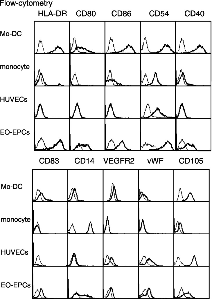
Fig. 3.
Expression of antigen-presenting molecules on EO-EPCs. Flow-cytometric analysis revealed the expression of HLA-DR, CD40, CD54, CD80, and CD86 on EO-EPCs, which are the molecules expressed on Mo-DCs. The DC marker, CD83, was also present on EO-DCs. CD14 is highly expressed on peripheral blood monocytes, and weakly on Mo-DCs, but EO-EPCs could be divided into the CD14(+) and (−) subpopulations. Although weaker than HUVECs, EO-EPCs expressed endothelial cell markers (VEGRF2, vWF, and CD105)

