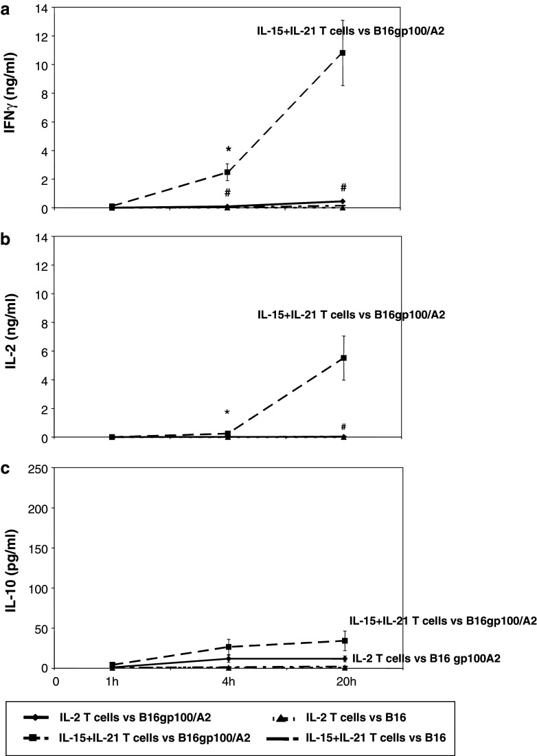
Fig. 5.
Antigen-specific secretion of IFNγ and IL-2, but not IL-10, shows accelerated kinetics upon combined treatment with IL-15 and IL-21. Murine splenocytes were gp100/A2 TCR-transduced and cultured with cytokines as described in legend to Fig. 1. Cytokine cultured T cells were stimulated with B16gp100/A2 and B16 cells for 1, 4 or 20 h, and analysed by commercial ELISA for the secretion of IFNγ (a), IL-2 (b) or IL-10 (c). Mock-transduced T cells showed no cytokine secretion at any time-point tested (data not shown). Time points following target cell stimulations are indicated at the X-axes. Absolute levels of cytokines present in supernatants are indicated at the Y-axes (mean ± SEM, n = 3, *,# p < 0.05 compared to IL-2 for B16gp100/A2 and B16, respectively)

