Fig. 5.
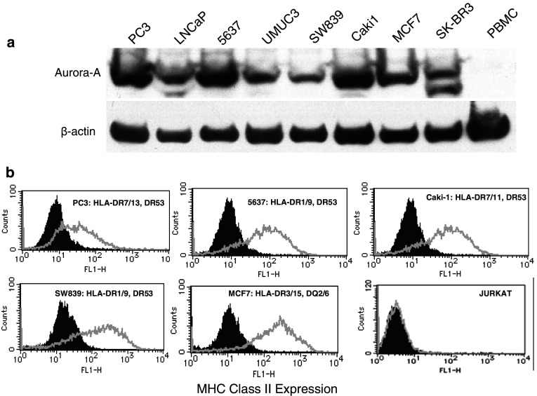
Evaluation of the expression of Aurora-A protein and cell surface MHC class II molecules by various human tumor cell lines. a Western blot analysis was done using an Aurora-A-specific mAb and β-actin-specific mAb as the control as described in “Materials and methods”. The Aurora-A protein has a mass of approximately 46 kDa. b Expression of cell surface MHC class II molecules on tumor cells evaluated by flow cytometry using anti-HLA class II mAb Tu39 conjugated with fluorescein isothiocyanate (thick line open histograms). Staining with the isotype-negative control (filled histograms). For each tumor, the previously determined MHC class II typing is shown

