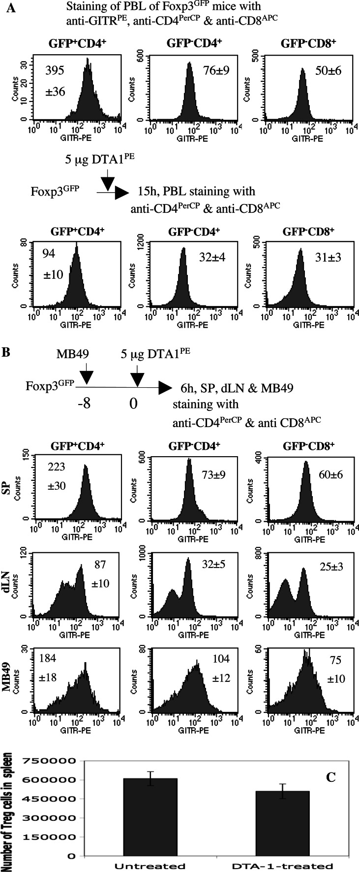
Fig. 4.
Treg cells constitutively express higher levels of GITR than conventional CD4 T cells in vivo. a Treg cells are primary targets of DTA-1. Top panels, the PBL from a group of Foxp3/GFP knock-in mice (n = 3) were stained with anti-GITRPE, anti-CD4PerCP and anti-CD8APC. The expression of GITR by GFP+CD4+, GFP−CD4+ and GFP−CD8+ T cells from individual mice was presented as histograms. MFI of GITR is shown as mean ± SEM. Bottom panels, a group of Foxp3/GFP knock-in mice were given 5 μg of anti-GITRPE (clone DTA-1) by i.v. injection. 15 h later, the PBL from individual mice were stained with anti-CD4PerCP and anti-CD8APC. MFI of GITR by GFP+CD4+, GFP−CD4+ and GFP−CD8+ T cells is presented as mean ± SEM. One representative of three independent experiments is shown. b Tumour-infiltrating Treg, CD4 and CD8 T cells express higher levels of GITR than those in dLN. A group of Foxp3/GFP knock-in mice (n = 4) were s.c. inoculated with MB49 cells (5 × 10⁵/mouse) 8 days previously. On day 0, the mice were i.v. injected with 5 μg of DTA-1PE. 6 h later, spleen, dLN and MB49 tumour cells from individual mice were stained with anti-CD4PerCP and anti-CD8APC. MFI of GITR by GFP+CD4+, GFP−CD4+ and GFP−CD8+ T cells is presented as mean ± SEM. One representative of two independent experiments is shown. c The number of splenic Treg cells was moderately reduced following DTA-1 treatment. Two groups of Foxp3/GFP knock-in mice (4 mice/group) were s.c. inoculated with 5 × 105 MB49 cells on day −8. The mice were untreated or treated with 50 μg of DTA-1 by i.p. injection on day 0. 3 days later, MB49 tumours from individual mice in each group were stained with anti-CD4PerCP. The absolute spleen-resident Treg cell numbers were determined by total spleen cells × % of lymphocyte gate × % of CD4+GFP+ cells within gated lymphocytes

