
Fig. 4.
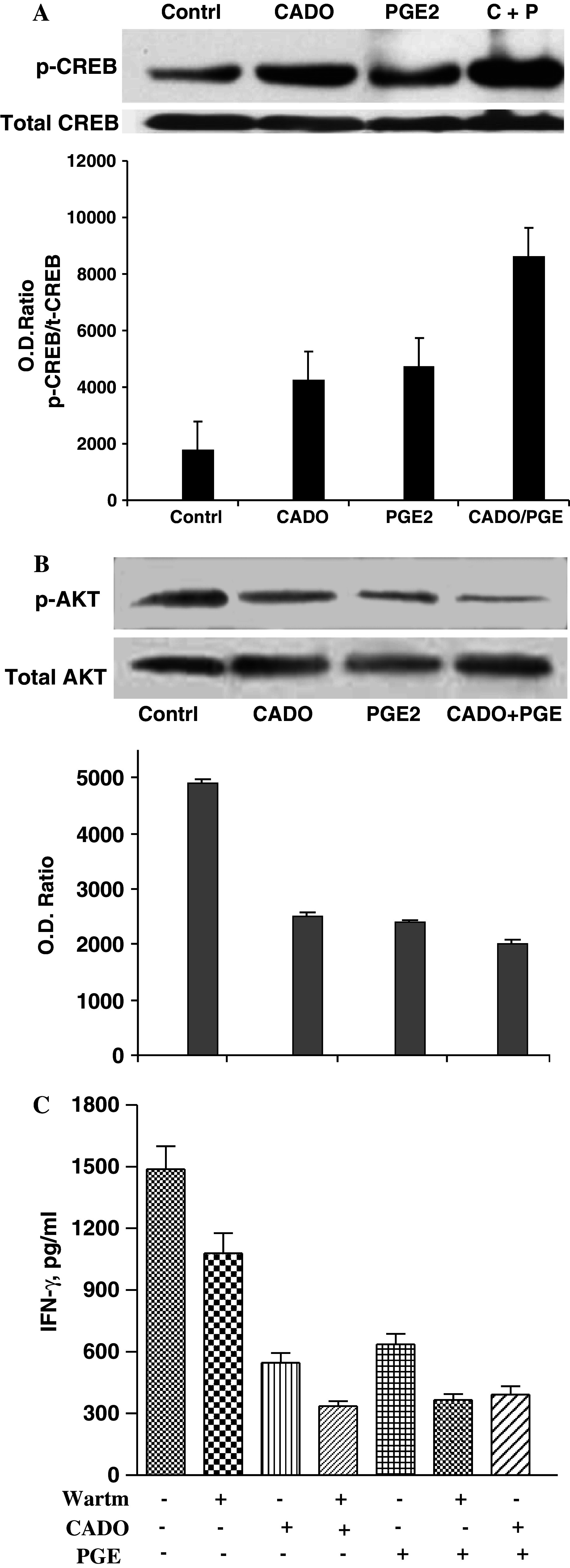
Western blot analysis of the effects of CADO and/or PGE2 on CREB (a) or Akt (b) phosphorylation. a LAK cells were incubated for 30 min with CADO (5 μM) and/or PGE2 (50 nM). Protein extracts (50 μg) were resolved by 10% SDS-PAGE and transferred to PVDF membranes, and the levels of total and phosphorylated CREB were assessed using specific antibodies. Corresponding densitometric analysis of optic density ratios between pCREB and total CREB is presented. b Effect of CADO and/or PGE2 on AKT phosphorylation. LAK cells were rested overnight without IL-2 and then were incubated with IL-2 (5,000 IU/ml) in the presence of CADO (25 μM) and/or PGE2 (500 nM) for 30 min. Protein extracts (50 μg) were prepared and resolved by 10% SDS-PAGE and transferred to PVDF membranes. The levels of total and phosphorylated AKT were assessed using specific antibodies. Corresponding densitometric analysis of optic density ratios between p-AKT and total AKT is presented. In rested LAK cells AKT was not phosphorylated (data not shown). c Wortmanin, an inhibitor of Akt phosphorylation poteniates the inhibitory effects of CADO and PGE2. The cytotoxic activity of LAK cells against 3LL tumor cells (E:T ratio 30:1) was tested in the presence of wortmanin (25 μM), CADO (5 μM) or PGE2 (6 nM). All groups significantly (P < 0.01) differ from control. The combined inhibitory effects in all groups were significantly (P < 0.05) higher than when each agent tested separately