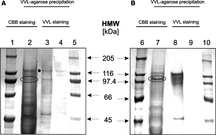
Fig. 2.
Lectin precipitation of Tn antigen-bearing glycoproteins in primary and metastatic melanoma. Extracts of WM793 (a) and Ma-Mel-27 (b) cellular proteins were precipitated using VVL-agarose, separated by SDS-PAGE and stained with CBB (lanes 2 and 7) or electrotransferred on PVDF membrane and probed with VVL (lanes 3 and 8) or VVL pre-blocked with GalNAc (lanes 4 and 9). In parallel, protein standards were resolved (lanes 1, 5, 6 and 10). Bands of interest (103 kDa for WM793 cell extract and 105 kDa for Ma-Mel-27 cell extract) were excised from the gel and subjected to mass spectrometry analysis (the encircled bands in lines 2 and 7). Note. One non-specific band was observed and marked with a black dot

