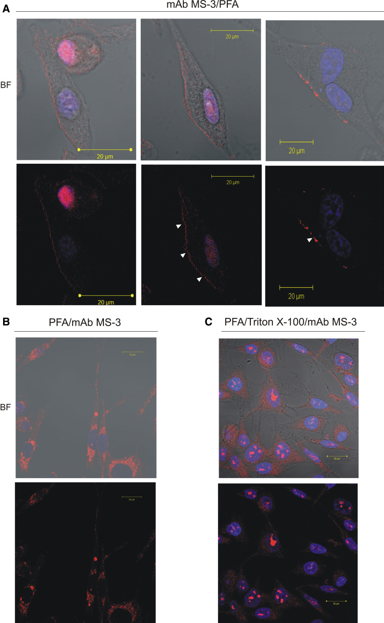
Fig. 5.
Detection of membrane-associated and intracellular nucleolin in melanoma cells by confocal immunofluorescence laser microscopy. For the detection of the cell-surface-clustered nucleolin (a), Ma-Mel-27 cells cultured in fresh medium for 5 h were further incubated for 1 h at 37°C in the presence of mAb MS-3 diluted 1:25 in 10% NGS, 2% BSA/PBS containing 25 mM NaN3 before PFA fixation. For staining of semi-permeabilised (b) or permeabilised cells (c) Ma-Mel-27 cells (after 5 h of culture in fresh medium) were fixed with 3% PFA or 2% PFA/Triton before incubation with mAb MS-3 in 1% BSA/PBS (overnight, RT) diluted 1:100, respectively. The bound anti-nucleolin antibody was revealed by Cy3-labeled goat anti-mouse antibodies (red). Nuclei were counterstained with DAPI (blue). BF; bright field. Note. In PFA/Triton-fixed cells, nucleolin was detected primarily as fine dot-like structures in the nuclei and also as spots in the cytoplasm (c). In cells preincubated with anti-nucleolin mAb (living unpermabilised cells) red patches indicate cell surface nucleolin (arrows in a) (color figure online)

