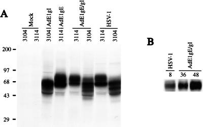
FIG. 2.
Expression of gE and gI by recombinant Ad vectors. (A) Human HEC-1A epithelial cells were left uninfected (mock) or were infected with 400 PFU of either Ad(E1−)gE or Ad(E1−)gI per cell, with both Ad(E1−)gE and Ad(E1−)gI, each at 400 PFU/cell, or with HSV-1 at 20 PFU/cell. The cells were labelled with [35S]methionine and [35S]cysteine for 3 h, beginning 48 h after infection with Ad vector or 8 h after infection with HSV. Detergent extracts of the cells were mixed with MAbs specific for gE (3114) or gI (3104) to immunoprecipitate these proteins. Positions of molecular size markers in kilodaltons are shown on the left. (B) HEC-1A cells were either infected with HSV-1 or coinfected with Ad(E1−)gE and Ad(E1−)gI. After 8 h of infection with HSV-1 or after 36 or 48 h of infection with the Ad vectors, the cell monolayers were scraped into SDS gel electrophoresis buffer. Samples were boiled, and the proteins were separated by polyacrylamide gel electrophoresis and then transferred to nylon membranes. gE was detected by incubating the blots with MAb II-481. The blots were washed and then incubated with horseradish peroxidase-coupled anti-mouse antibodies, and these antibodies were detected by enhanced chemiluminescence.

