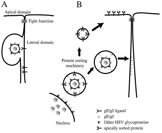
FIG. 7.
Models for how gE-gI facilitates epithelial cell-to-cell spread. (A) gE-gI (triangles) accumulates at junctions formed between an infected cell (left) and an uninfected cell (right) by virtue of interactions with cellular ligands which are expressed in the lateral membranes of the uninfected cell. Presumably, binding of gE-gI to these cellular ligands enhances the transfer of virus across cell junctions so that virus particles can move into the space between the cells and then fuse with the opposing, uninfected cell membrane. It is not clear whether gE-gI functions in this capacity as part of the virion envelope (top) or as a constituent of the plasma membrane of the infected cell (bottom). (B) Alternatively, gE-gI may act as a trafficking signal to direct enveloped particles to the lateral plasma membrane rather than to the apical surface (as with apically sorted proteins). Since there is some evidence that the cytoplasmic domains of gE and gI have motifs that affect intracellular transport, it is likely that these cytosolic domains serve to target virions present within cytoplasmic vesicles to sites of cell-cell contact.

