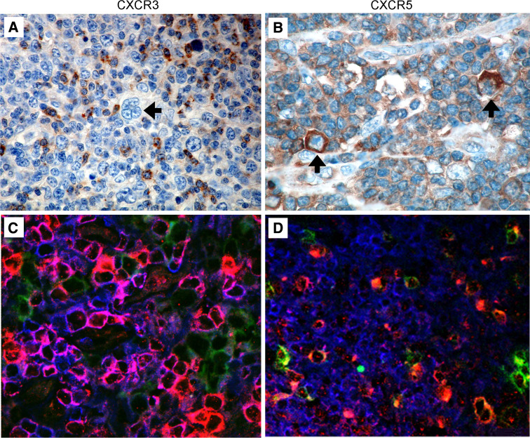
Fig. 2.
Expression of CXCR3 and CXCR5 in HL tissue. CXCR3 and CXCR5 chemokine receptor expression was examined by immunohistochemical staining of formalin-fixed HL biopsies (n = 9). a CXCR3+ lymphoid cells were detected in the vicinity of cells having H-RS morphology. b CXCR5 was expressed on a minor population of lymphoid cells as well as H-RS cells. H-RS cells are indicated with an arrow. CXCR3 (c) and CXCR5 (d) expression was further investigated with triple immunofluorescent staining of snap frozen HL biopsies (n = 8) for CD3 (blue), CD30 (green) and CXCR3 or CXCR5 (red). Individual images were merged to provide a composite with CXCR3+ or CXCR5+ T cells staining pink

