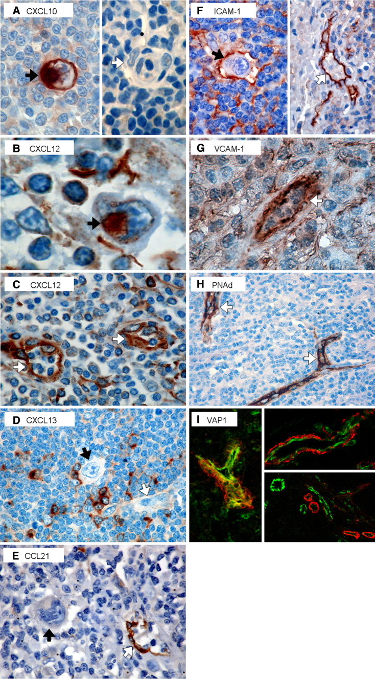
Fig. 3.
Chemokine and adhesion molecule expression in HL. HL cases (n = 9) were examined by immunohistochemistry for expression of the following chemokines and adhesion molecules: CXCL10 (a), CXCL12 (b, c), CXCL13 (d), CCL21 (e), ICAM-1 (f), VCAM-1 (g), and PNAd (h). H-RS cells and vessels are indicated with black and white arrows, respectively. VAP-1 expression on 8 HL biopsies was examined by immunofluorescence (i). Colocalisation of VAP-1 (red) with the vascular endothelium was investigated by co-staining with CD31 (green)

