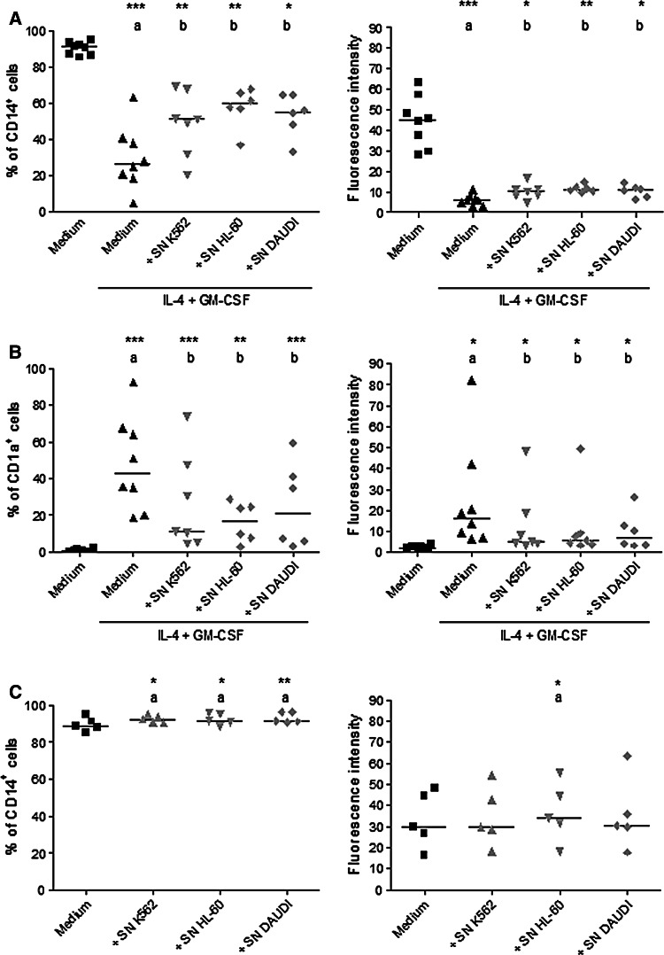
Fig. 2.
CD14 and CD1a expressions by monocytes after induction of differentiation in the presence of K562, HL-60 and DAUDI supernatants. Monocytes isolated from healthy volunteers were incubated with or without IL-4 and GM-CSF in the presence of K562, HL-60 and DAUDI supernatants (10% final volume). After 5 days, CD14 and CD1a expressions were analyzed by flow cytometry. Data show the percentage of cells stimulated with IL-4 and GM-CSF that express CD14 (left) and mean of fluorescence intensity of CD14 (right) (a); the percentage of cells stimulated with IL-4 and GM-CSF that express CD1a (left) and mean of fluorescence intensity of CD1a (right) (b); the percentage of cells not stimulated with IL-4 and GM-CSF that express CD14 (left) and mean of fluorescence intensity of CD14 (right) (c). Each symbol per group represents one individual assessed under the different conditions. The horizontal lines represent the median of at least five independent experiments. a Compared with monocytes without IL-4 and GM-CSF, b compared with monocytes cultured with IL-4 and GM-CSF. *Significantly different (P < 0.05), **Significantly different (P < 0.01), ***Significantly different (P < 0.001)

