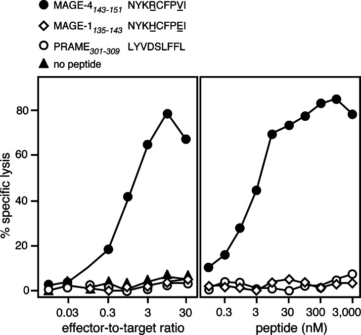
Fig. 2.
Lysis of an HLA–A24 target loaded with MAGE-4 peptide NYKRCFPVI. a HLA–A24 EBV-B cells from donor LB2348 were 51Cr-labeled for 1 h, incubated for 5 min with 1 μg/ml of one of the indicated peptides and incubated with CTL at indicated effector-to-target ratios. The MAGE-4 and the MAGE-1 peptide are very similar. The amino acid differences are underlined. Chromium release was measured after 4 h. b HLA–A24 EBV-B cells were 51Cr-labeled for 1 h, incubated for 15 min with threefold dilutions of each of the synthetic peptides. CTL was subsequently added at an effector-to-target ratio of 10. Chromium release was measured 4 h later. The concentration of peptide indicated in the figure corresponds to the concentrations during the 4 h of incubation after addition of the CTL

