
Fig. 3.
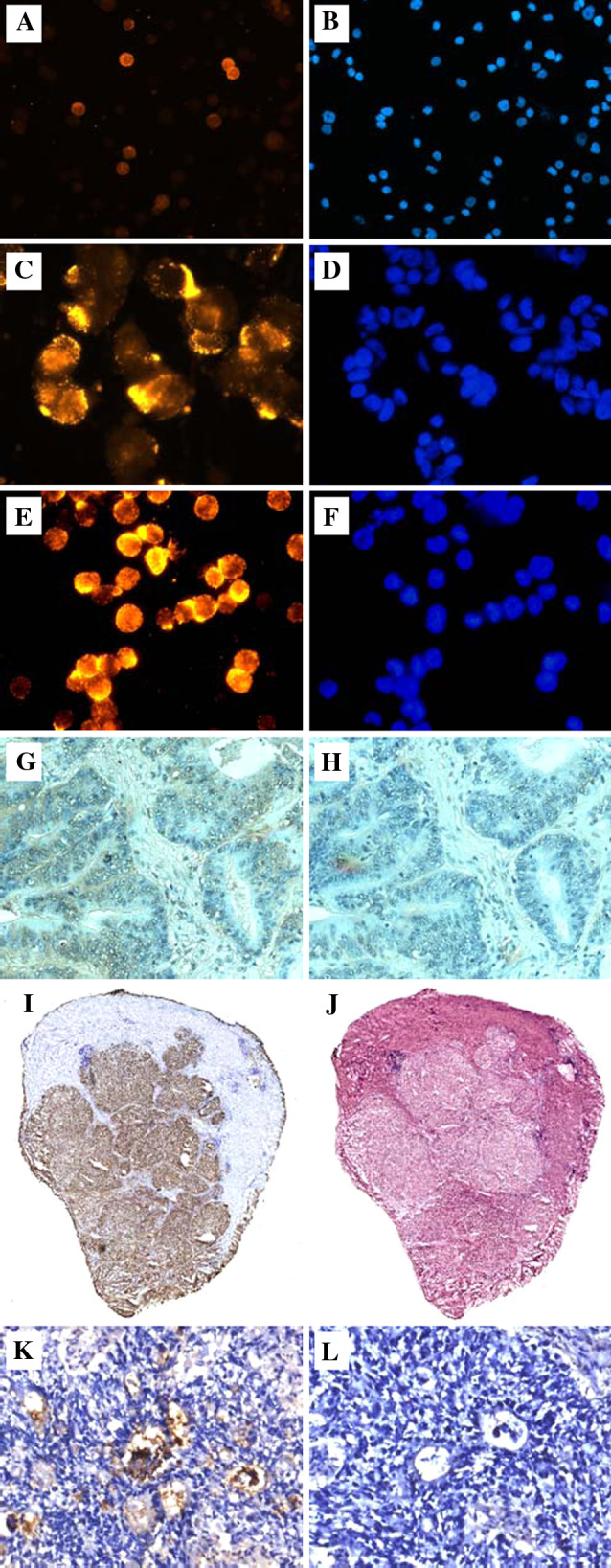
Immunostaining of human tumor cells and tissues with the trimeric anti-TF multibody scFv(1aa). a, b KG-1 (AML cell line); c, d ZR-75-1 (breast cancer cell line); e, f NM-D4 (glycoengineered CML cell line); a, c, e stained in immunofluorescence (IF); b, d, f counterstaining of nuclei with DAPI (identical fields as in IF). In a, around ten out of 80 cells (12.5%) are membrane-stained by the multibody with different intensities. This pattern is identical to that seen with the mouse anti-TF antibody A78-G/A7 (not shown). g Cryosection of human colon carcinoma stained in immunohistochemistry (IHC), h adjacent section to g as omission control. i Whole-size cryosection of human hepatocellular carcinoma stained in IHC, j adjacent section stained with hematoxylin/eosin. k cryosection of a primary human colon carcinoma transplanted s.c. into an NCR:nu/nu mouse stained in IHC, l omission control