Fig. 3.
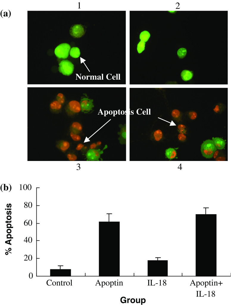
LLC cell death induced by pIL-18 and pAPOPTIN. a Cells at 1 × 10⁶ cells/well were transfected with pIL-18 and pAPOPTIN for 48 h, stained with AO/EB, and their morphology was assessed immediately using florescence microscopy (×400). (1) Control group: viable cells with green nuclei and intact structure. (2) pIL-18 group: pIL-18 treatment for 48 h. There were no visible apoptosis-associated morphological alternations in LLC cells. (3) pAPOPTIN group: pAPOPTIN treatment for 48 h. Cells in late apoptosis were present, characterized by dense orange condensation of chromatin in nuclei and reduced cell size (arrows). (4) pIL-18 + pAPOPTIN group: pIL-18 + pAPOPTIN treatment for 48 h. As above, cells in late apoptosis were present, characterized by dense orange condensation of chromatin in nuclei and reduced cell size (arrows). b This procedure was used to quantify the number of apoptotic cells after treatment with pIL-18 and pAPOPTIN

