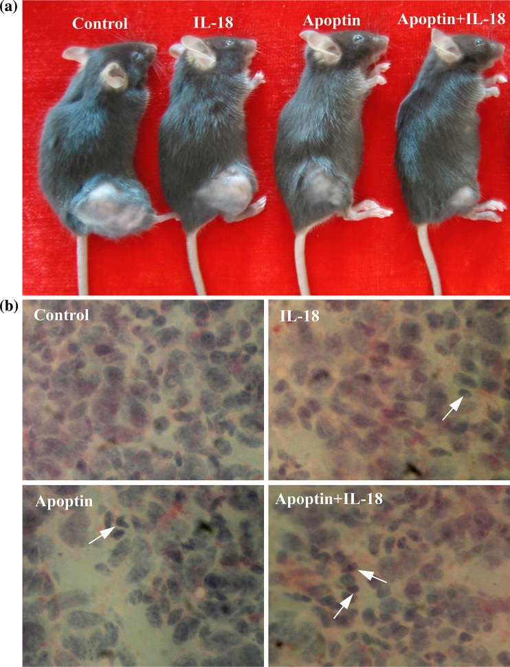
Fig. 5.
Macroscopic and microscopic appearances after treatment of C57BL/6 mice bearing LLC. a Macroscopic appearance in control and pIL-18 and/or pAPOPTIN-treated mice. Note the significant decrease in tumor volume associated with pIL-18 + pAPOPTIN treatment. b Hematoxylin–eosin staining of the tumor from control and pIL-18 and/or pAPOPTIN-treated mice. Note the significant aberrant morphology characterized by loss of tissue integrity, increase in interstitial space, and visible remnants of disintegrated cells in tumor areas after Apoptin and IL-18 treatment. Arrows indicate tumor cells containing condensed dark nuclei. (original magnification: ×400)

