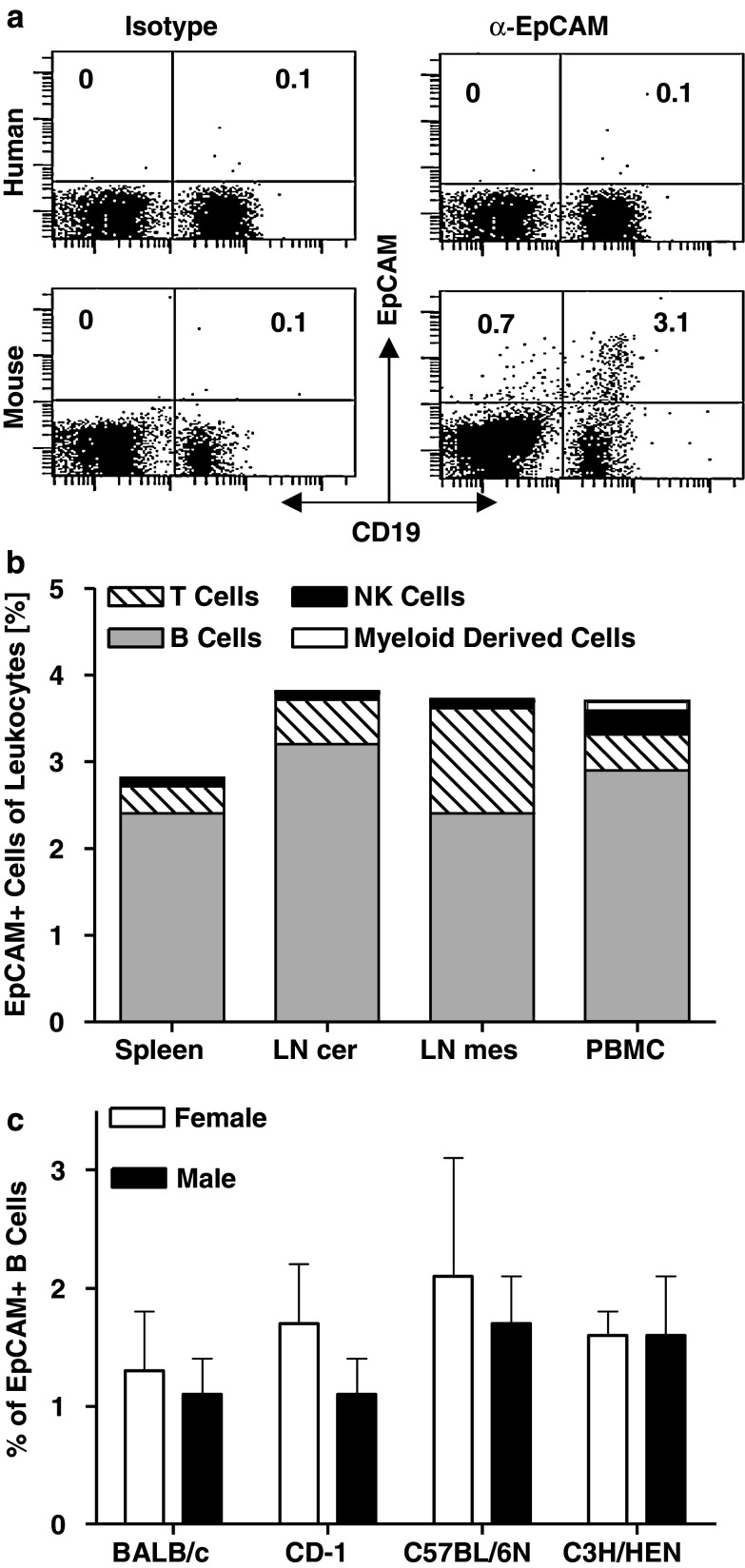
Fig. 4.
EpCAM+ cells are present in blood and lymph nodes of mice. a Human PBMC (upper panel) and murine splenocytes (lower panel) were double-stained for CD19 and EpCAM, or with corresponding isotype control antibodies. b Cells isolated from spleen, cervical (LN cer) and mesenteric lymph nodes (LN mes) as well as from peripheral blood (PBMC) of two male BALB/c mice were stained for CD3 (T cells), CD19 (B cells), CD49b (NK cells) and Ly6G (myeloid derived cells) in combination with an anti-murine EpCAM mAb, or the corresponding isotype control. Percentage of cells positive for EpCAM and the respective markers are shown. c PBMC of six female and male BALB/c, C57BL/6 N, C3H/HEN and CD-1 mice were stained for CD19 and EpCAM and analyzed by flow cytometry. Sex and strain specific percentage of CD19/EpCAM double positive cells are shown. Error bars indicate standard error of the mean

