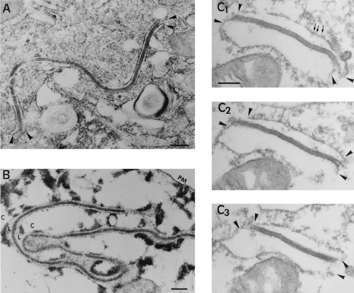
FIG. 5.
Topology of the zipper-like structures. (A and B) Ultrathin Epon sections of marginal zipper-like structures within a nonpermeabilized (A) or SLO-permeabilized (B) infected cell at 16 h p.i. Note in the control (A) how the limits of the viral intermediate appear to be continuous with two adjacent ER cellular cisternae (arrowheads). After cell permeation (B), the cytosol (C) is extracted but not the luminal contents (L), which become obvious. Thus, it is clear that the core shell is a cytosolic structure limited by two ER cisternae. (C1 to C3) Set of serial sections of a peripheral zipper-like structure. The core shell is interpreted as a laminar structure whose limiting surfaces interact with the cytoplasmic sides of ER cisternae. Note the presence of ribosomes (small arrows) attached to ER membranes (arrowheads). Bars: 200 nm (A and B) and 100 nm (C1).

